Shoulder Anatomy Diagram : Anatomy And Injuries Of The Shoulder Anatomical Chart Shoulder Injury / Learn about these muscles, their origin and insertion points, and their functional anatomy.
Shoulder Anatomy Diagram : Anatomy And Injuries Of The Shoulder Anatomical Chart Shoulder Injury / Learn about these muscles, their origin and insertion points, and their functional anatomy.. Arthritis and joint pain arthritis and joint pain infographic, anatomic illustration of an inflammed hand and arm shoulder anatomy stock. The shoulder muscles consist of the deltoids and the rotator cuff group. Shoulder joint the most flexible joint in the entire human body, our shoulder joint is formed by the union of the humerus, the scapula (or shoulder blade), and the clavicle (or collarbone). The muscles of the shoulder support and produce the movements of the shoulder girdle.they attach the appendicular skeleton of the upper limb to the axial skeleton of the trunk. Shoulder muscle anatomy shoulder blade muscles body muscle anatomy human body anatomy anatomy male arm anatomy medical anatomy anatomy drawing muscle diagram.
Numerous muscles help stabilize the three joints of. Shoulderdoc.co.uk satisfies the intute criteria for quality and has been awarded 'editor's choice'. Arthritis and joint pain arthritis and joint pain infographic, anatomic illustration of an inflammed hand and arm shoulder anatomy stock. The acromioclavicular joint is where the acromion, part of the shoulder blade (scapula) and the collar bone (clavicle) meet. Another name for this bone is the shoulder blade.
1 from Learn their origins/insertions, functions & exercises. This page is about shoulder tendon anatomy diagram,contains muscles attachment of rotator cuff muscle,upper extremity occupational therapy 205 with teresa at tufts university,soft tissues of the shoulder,biceps and triceps tendon rupture and more. Shoulderdoc.co.uk satisfies the intute criteria for quality and has been awarded 'editor's choice'. Here is an overview of the shoulder bones: It is the major joint connecting the upper limb to the trunk. Shoulder joint anatomy and pain. Three bones come together at the shoulder joint. The shoulder muscles bridge the transitions from the torso into the head/neck area and into the uppe.
The shoulder is made up of two joints, the acromioclavicular joint and the glenohumeral joint.
The shoulder is a complex combination of bones and joints where many muscles act to provide the widest range of motion of any part of the body. There are many nerves and blood vessels that supply the muscles and bones of the shoulder. Shoulder osteoarthritis (oa) may be a significant cause of pain and disability. The list of muscles and their functions are presented below. Related posts of diagram of shoulder muscles and tendons muscle anatomy multiple choice. The glenohumeral joint is where the ball (humeral head) and the socket (the glenoid) meet. The latissimus dorsi and teres major on the posterior. On the anterior side of the shoulder, the coracobrachialis, serratus anterior, pectoralis major, and pectoralis minor muscles work as a group to flex and adduct the scapula and humerus anteriorly toward the sternum. Development of boney growths, called osteophytes or bone spurs, may impede this function and cause pain. Subscapularis, supraspinatus, infraspinatus and teres minor. Another name for this bone is the shoulder blade. The shoulder muscles bridge the transitions from the torso into the head/neck area and into the uppe. 3 problems of the shoulder.
The glenohumeral joint is where the ball (humeral head) and the socket (the glenoid) meet. You will receive your score and answers at the end. Choose an answer and hit 'next'. Much of your shoulder motion is between the. Nerves are like electrical wires that carry signals from the brain to the muscles to allow for movement of.
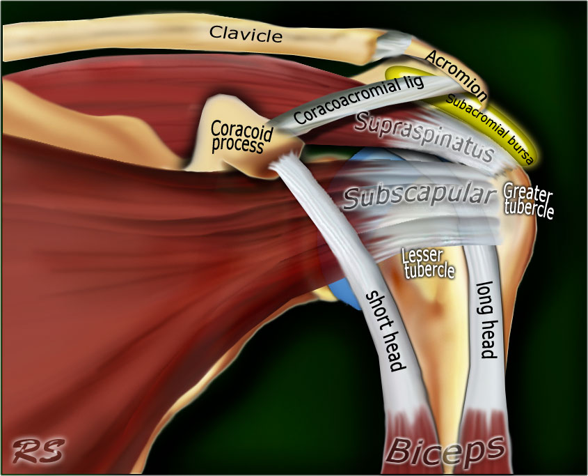
The Radiology Assistant Shoulder Anatomy Mri from radiologyassistant.nl And you can see superiorly, this is the tendon of the long head of the biceps and it's been cut. Nerves are like electrical wires that carry signals from the brain to the muscles to allow for movement of. August 2, 2017 / the hand society. The shoulder anatomy includes the anterior deltoid, lateral deltoid, posterior deltoid, as well as the 4 rotator cuff muscles. This site complies with the honcode standard for trustworthy health information: Muscles of the shoulder : The bones must maintain their strength and smooth surface in order to move easily against each other. It is one of the most mobile joints in the human body, at the cost of joint stability.
Several ligaments make up parts of the joint capsule, and these ligaments are important in keeping the shoulder joint in proper position.
I've just switched over to this diagram here and we're looking at the same view, a lateral view of the right shoulder. Hooked acromions are the third type of acromion. The main shoulder muscles are trapezius, deltoid, pectoralis major and 4 rotator cuff muscles: Ebraheim's educational animated video describes muscle anatomy of the shoulder girdle and anatomy of the shoulder joint.anatomy of the shoulder muscles a. The anatomy of the shoulder. There are five major bones in the shoulder. The coordinated activity of numerous muscles working together in set patterns is required to produce this motion ; Shoulder joint the most flexible joint in the entire human body, our shoulder joint is formed by the union of the humerus, the scapula (or shoulder blade), and the clavicle (or collarbone). Starting with what is deepest, it goes: Discuss tha agaonist/antagonist relationship of muscles. Labeled anatomy chart of male triceps and back muscles on white background labeled human anatomy diagram of man's arm, shoulder and upper back muscles in a posterior view on a white background. Name this muscle that elevates the shoulder. An image depicting shoulder anatomy can be seen below.
It is made up of four joints and five. The coordinated activity of numerous muscles working together in set patterns is required to produce this motion ; The bones of the pectoral girdle (clavicle and scapula) provide increased mobility to the shoulder joint by allowing it to move in all directions. Three bones come together at the shoulder joint. The shoulder joint is protected superiorly by an arch, which is formed by the coracoid process of the scapula, the acromion process of the scapula and the clavicle.
Shoulder Injury Hurt911 from hurt911.org The capsule separates the joint from the rest of the body and contains the joint fluid. Numerous muscles help stabilize the three joints of. You will receive your score and answers at the end. Much of your shoulder motion is between the. Development of boney growths, called osteophytes or bone spurs, may impede this function and cause pain. Several ligaments make up parts of the joint capsule, and these ligaments are important in keeping the shoulder joint in proper position. The bones of the pectoral girdle (clavicle and scapula) provide increased mobility to the shoulder joint by allowing it to move in all directions. Shoulderdoc.co.uk satisfies the intute criteria for quality and has been awarded 'editor's choice'.
This diagram depicts picture of shoulder muscles with parts and labels.
Here i have explained the shoulder muscles and in my previous video i have talked about the shoulder j. Discuss tha agaonist/antagonist relationship of muscles. The shoulder is located where the arm meets the torso and is comprised of and functions with the following basic components: August 2, 2017 / the hand society. Name this muscle that elevates the shoulder. To keep things simple, we can divide the shoulder into layers. Several ligaments make up parts of the joint capsule, and these ligaments are important in keeping the shoulder joint in proper position. Shoulderdoc.co.uk satisfies the intute criteria for quality and has been awarded 'editor's choice'. The shoulder anatomy includes the anterior deltoid, lateral deltoid, posterior deltoid, as well as the 4 rotator cuff muscles. Shoulder muscle anatomy shoulder blade muscles body muscle anatomy human body anatomy anatomy male arm anatomy medical anatomy anatomy drawing muscle diagram. You can see this fibrocartilaginous collar, the glenoid labrum surrounding the glenoid fossa. This diagram depicts picture of shoulder muscles with parts and labels. We've got the acromion posteriorly and the coracoid process anteriorly.


Komentar
Posting Komentar